The lateral pectoral nerve is larger than the medial and arises from the upper and middle trunks or by a single branch from the lateral cord of the brachial plexus. Its fibers are derived from the fifth to seventh cervical rami (Gray’s Anatomy, 1995). It crosses anterior to the axillary artery and vein, pierces the clavipectoral fascia, and supplies the deep surface of the pectoralis major. It sends a small branch to the medial pectoral nerve, forming a loop in front of the first part of the axillary artery, to supply fibers of the pectoralis minor. The medial pectoral nerve is derived from the eighth cervical and first thoracic cervical rami. It arises from the medial cord of the brachial plexus, posterior to the axillary artery. It curves forward to join the branch from the lateral pectoral nerve, entering the deep surface of the pectoralis minor to supply it. Branches from the medial pectoral nerve may also supply portions of the pectoralis major.
Anatomical Illustrations
Pectoralis Major
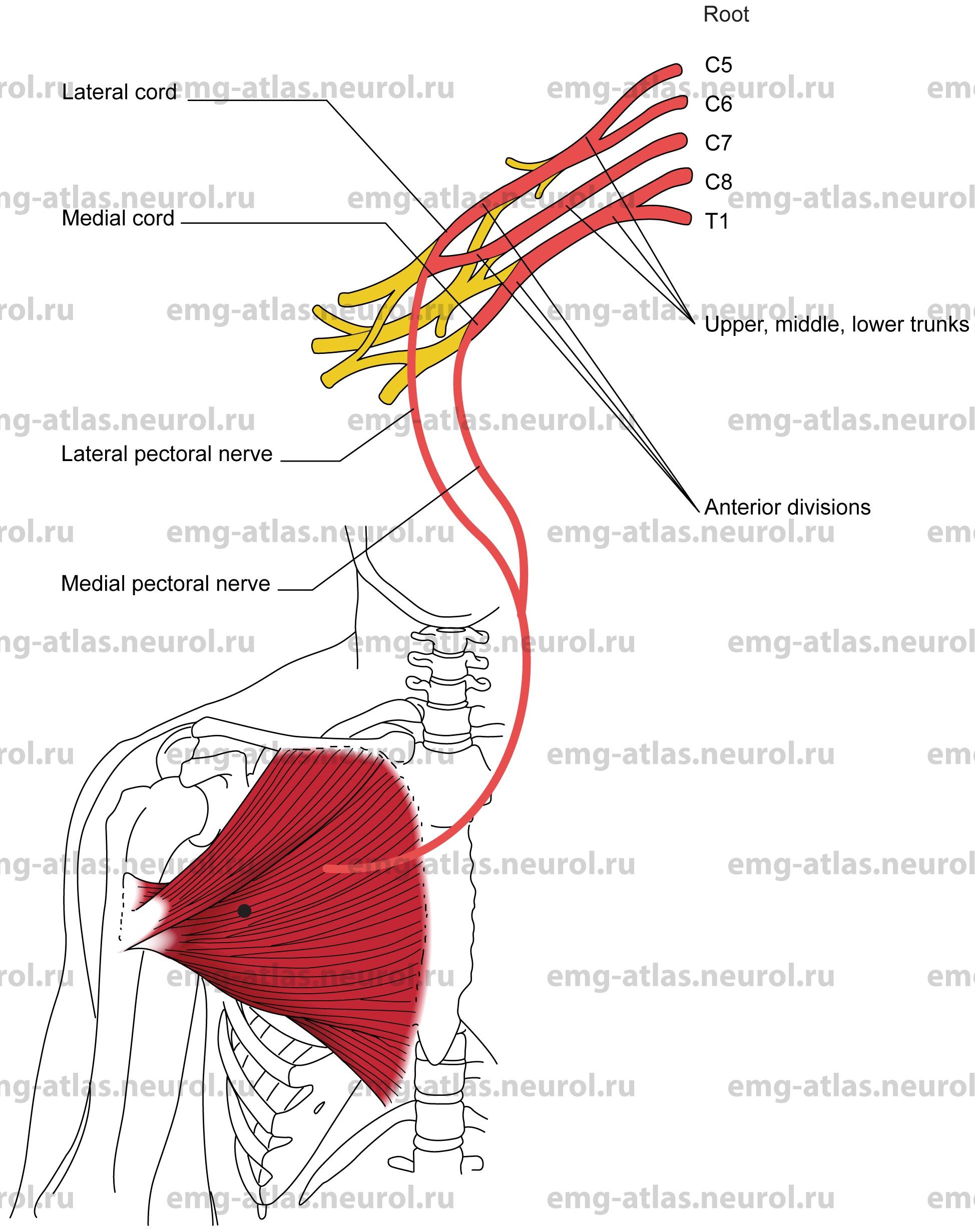
Innervation
Clavicular part: Innervation is via the lateral pectoral nerve, lateral cord, upper trunk, and roots C5, C6.
Sternocostal part: Innervation is via the lateral and medial pectoral nerves, lateral and medial cords, middle and lower trunks, and roots C7, C8, T 1.
Origin
The clavicular part originates at the sternal half of the clavicle.
The Sternocostal part originates at the anterior surface of the sternum, the cartilage of the first six or seven ribs, and the aponeurosis of the external oblique muscle of the abdomen.
Insertion
Insertion is at the lateral lip of the intertubercular sulcus on the shaft of the humerus.
Activation Maneuver
Adduction of the arm activates the muscle.
EMG Needle Insertion
Insert the needle just medial to the anterior axillary fold over the bulk of the muscle.
Pitfalls
If the needle is inserted too superiorly, it may be in the anterior fibers of the deltoid, which is supplied by the axillary nerve.
If the needle is inserted too laterally, it may be in the coracobrachialis or the short head of the biceps, which are supplied by the musculocutaneous nerve.
Clinical Comments
An isolated lesion of the lateral or medial pectoral nerves is rare.
Weakness of the pectoralis major results in limited adduction of the arm.
Neurogenic EMG changes in the pectoralis major may be seen with radiculopathies affecting the C5-T 1 roots. This muscle is therefore of little benefit in localizing a root lesion.
Anatomical Illustrations
Pectoralis Minor
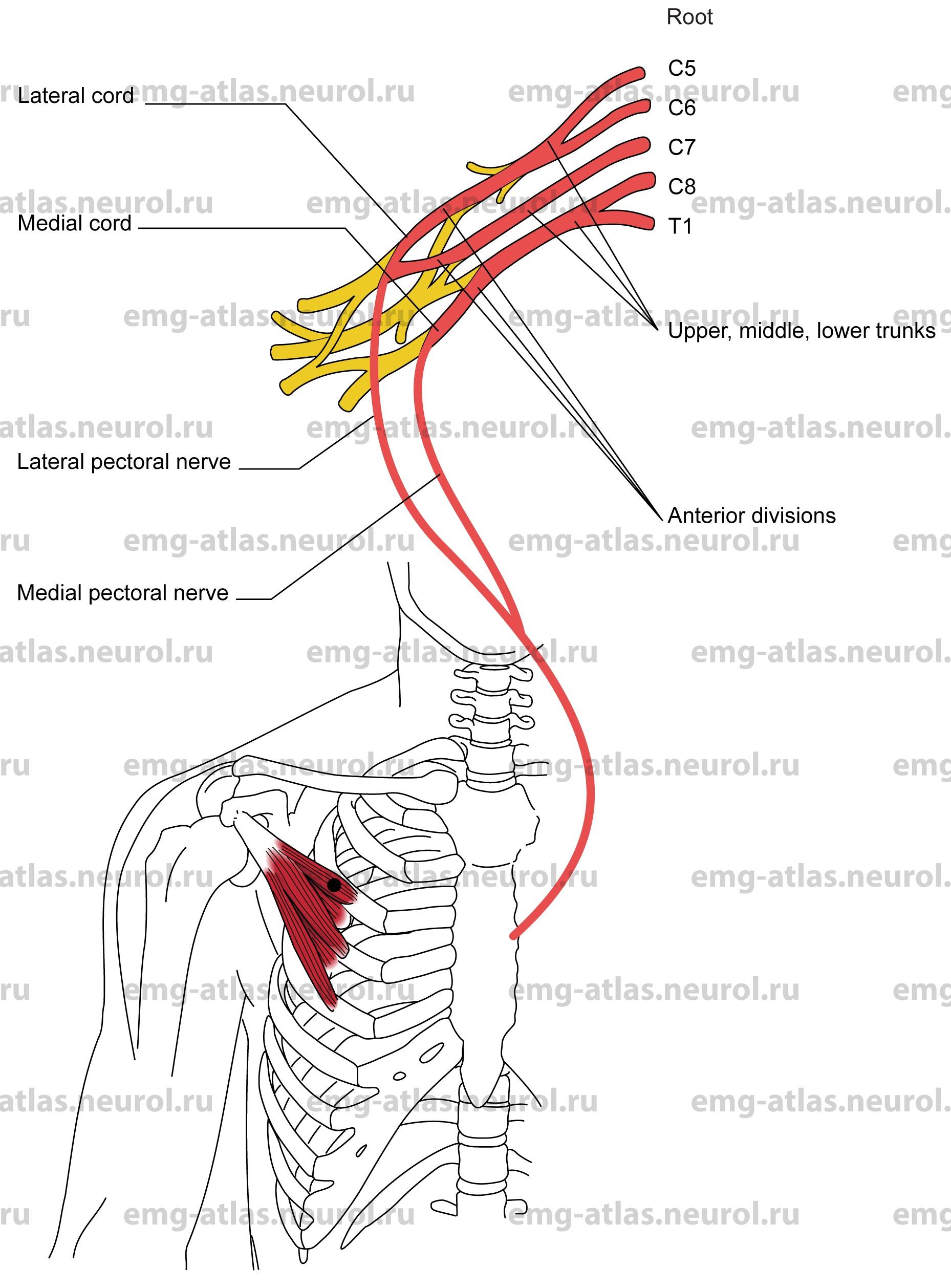
Innervation
Innervation is via the medial and lateral pectoral nerves; medial and lateral cords; upper, middle, and lower trunks; and roots C5, C6, C7, C8, and T1.
Origin
The pectoralis minor originates at the outer surfaces of the third to fifth ribs (frequently second to fourth).
Insertion
Insertion is at the coracoid process of the scapula.
Activation Maneuver
Depression of the shoulder activates the muscle.
EMG Needle Insertion
Insert the needle in the midclavicular line overlying the third rib
Pitfalls
If the needle is inserted too superficially, it will be in the pectoralis major.
Clinical Comments
An isolated lesion of the lateral or medial pectoral nerves is rare.
Weakness of the pectoralis minor is unlikely to be clinically detectable.
Neurogenic EMG changes in the pectoralis minor may be seen with radiculopathies affecting the C5-T, roots. This muscle is therefore of little benefit in localizing a root lesion.
In prolonged hyperabduction of the arm, the neurovascular bundle in the axilla can be stretched under the pectoralis minor tendon, resulting in symptoms of neurovascular compression (Pecina et al., 1997).